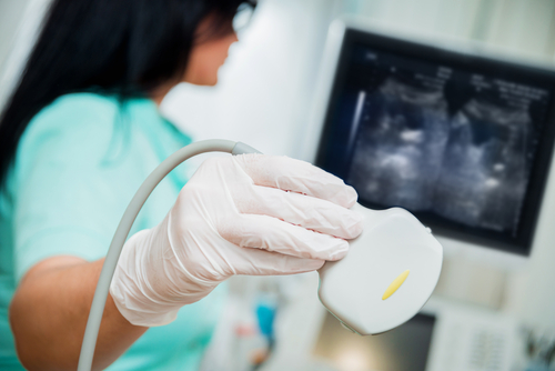
Ultrasound Imaging
An abdominal ultrasound image is a useful way of examining internal organs, including the liver, gallbladder, spleen, pancreas, kidneys, and bladder. Because US images are captured in real time, they can show movement of internal tissues and organs and enable physicians to see blood flow. This can help to diagnose a variety of conditions and to assess damage caused by illness.
Ultrasound imaging is used extensively for evaluating the kidneys, liver, gallbladder, pancreas, spleen and blood vessels of the abdomen. Because it provides real-time images, it can also be used to:
Guide procedures such as needle biopsies, in which needles are used to sample cells from organs for laboratory testing
Help a physician determine the source of many abdominal pains, such as stones in the gallbladder or kidney, or an inflamed appendix
Help identify the cause for enlargement of an abdominal organ
Doppler ultrasound is a special type of ultrasound study that examines major blood vessels. These images can help the physician to see and evaluate:
Blockages to blood flow, such as clots
Build-up of plaque inside the vessel
Conge
With knowledge about the speed and volume of blood flow gained from an ultrasound image, the physician can often determine whether a patient is a good candidate for a procedure like angioplasty
PELVIC ULTRASOUND IMAGING
Pelvic ultrasound (see right) is most often used to examine the uterus and ovaries and, during pregnancy, to monitor the health and development of the embryo or fetus. In men, a pelvic ultrasound usually focuses on the bladder and the prostate gland.OBSTETRIC ULTRASOUND
Obstetric ultrasound refers to the specialized use of sound waves to visualize and thus determine the condition of a pregnant woman and her embryo or fetus.CAROTID AND ABDOMINAL AORTA ULTRASOUND IMAGING
Ultrasound of the carotid arterial system provides a fat, noninvasive means of identifying blockages of blood flow in the neck arteries to the brain that might produce a stroke or mini-stroke. Ultrasound of the abdominal aorta is primarily used to evaluate for an aneurysm which is an abnormal enlargement of the aorta usually from atherosclerotic disease.VENOUS ULTRASOUND IMAGING
The most common reason for a venous ultrasound exam is to search for blood clots, especially in the veins of the leg. These clots may break off and pass into the lungs, where they can cause a dangerous condition called pulmonary embolism. If found in time there are treatments that can prevent this from happening.Venous Ultrasound Imaging is used for:
Finding the cause of long-standing leg swelling. In people with varicose veins, a common condition, the valves that keep blood flowing in the right direction may not work well, and venous ultrasound can help the surgeon decide how best to deal with this condition.
Aid placement of a needle or catheter in a large interior vein. Sonography can help locate the exact site of the vein and avoid complications such as bleeding or air in the chest cavity.
Map out the veins in the leg or arm so that segments may be removed and used to bypass an area of disease. An example is using pieces of vein from the leg to surgically bypass narrowed coronary arteries.
Examine a blood vessel graft used for dialysis if it is not working as expected: an area of narrowing in the graft may be responsible.