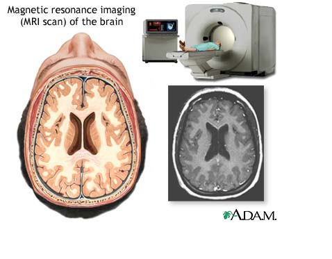
Magnetic Resonance Imaging (MRI)
What is an MRI of the Body?
Magnetic Resonance Imaging (MRI) is a noninvasive medical test that helps physicians diagnose and treat medical conditions.
MRI uses a powerful magnetic field, radio frequency pulses and a computer to produce detailed pictures of organs, soft tissues, bone and virtually all other internal body structures. The images can then be examined on a computer by a radiologist. The images can be stored and copied on a CD. The MRI does not use ionizing radiation (x-rays).
Detailed MR images allow physicians to better evaluate various parts of the body and certain diseases that may not be assessed adequately with other imaging methods such as x-ray, ultrasound or computed tomography.
MRI of the body is performed to evaluate:
Blood Vessels
Breasts
Organs of the chest and abdomen - including the heart, liver, biliary tract, kidney, spleen, and pancreas and adrenal glands
Pelvic organs including the reproductive organs in the male (prostate and testicles) and the female (uterus, cervix and ovaries)
Physicians use the MRI examination to help diagnose or monitor treatment for conditions such as:
Blockages, or enlargements of blood vessels, including the aorta, renal arteries, and arteries in the legs
Breast cancer and implants
Causes of pelvic pain in women, such as fibroids, endometriosis and adenomyosis
Certain types of heart problems
Cysts and solid tumors in the kidneys and other parts of the urinary tract
Diseases of the liver, such as cirrhosis and that of other abdominal organs, including the dile ducts, gallbladder, and pancreatic ducts
Suspected uterine congential abnormalities/anomalies in women undergoing evaluation for infertility
Tumors and other abnormalities of the reproductive organs (e.g. uterus, ovaries, testicles, prostate)
Tumors of the chest, abdomen, or pelvis